Connective Tissue
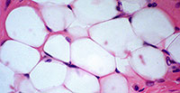
Adipose Tissue
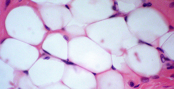
Structure
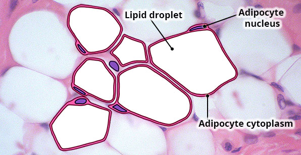
Consists of clusters of large adipocytes (fat cells); each adipocyte contains a large, centrally located lipid droplet that pushes the nucleus and cytoplasm to the edge of the cell.
Location
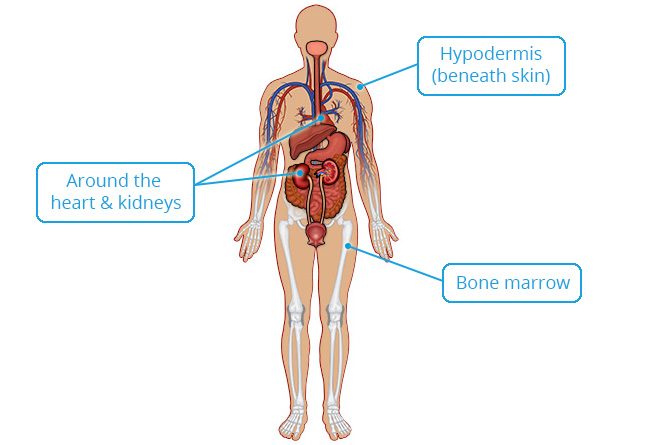
Subcutaneous layer (hypodermis) beneath skin; around heart and kidneys; yellow bone marrow; breast.
Function
Energy storage; reduces heat loss; protective cushioning for organs.
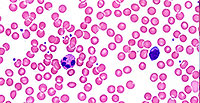
Blood Tissue
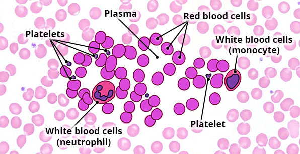
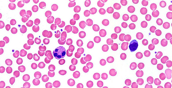
Structure
Mostly red blood cells (pink discs with no nuclei), fewer white blood cells, platelets and plasma.
Location
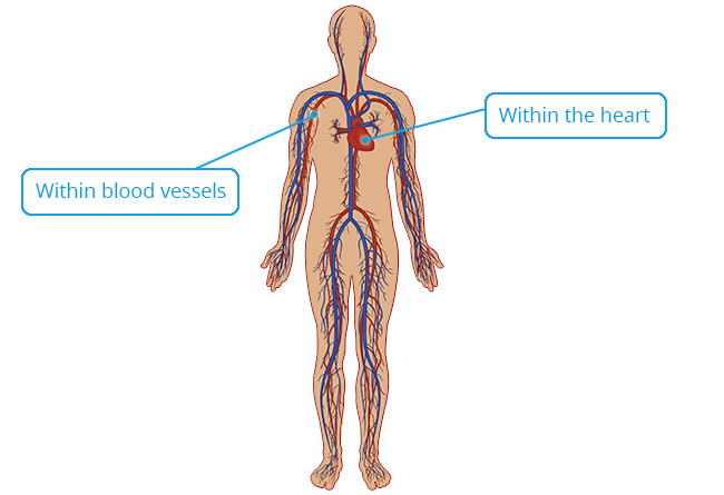
Within blood vessels and the heart.
Function
Red blood cells transport gases (oxygen and carbon dioxide); white blood cells protect the body from infections and are involved in allergic reactions; platelets help to clot blood; plasma transports nutrients, wastes and other substances throughout the body.
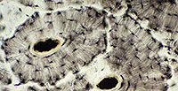
Bone Tissue
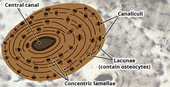
Structure
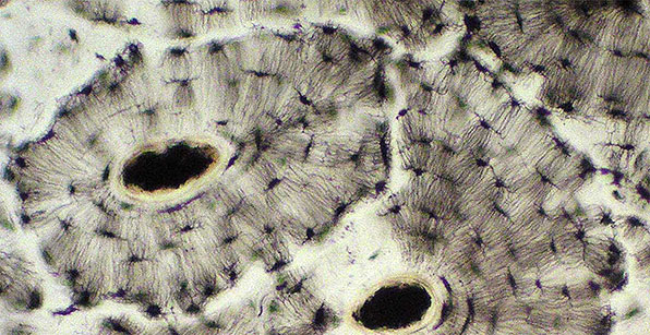
Hard, calcified bone matrix arranged in concentric rings (lamellae) around central (haversian) canals that contain blood vessels; osteocytes (not visible in this image of ground bone) reside in lacunae and connect to each other via tiny channels called canaliculi.
Location
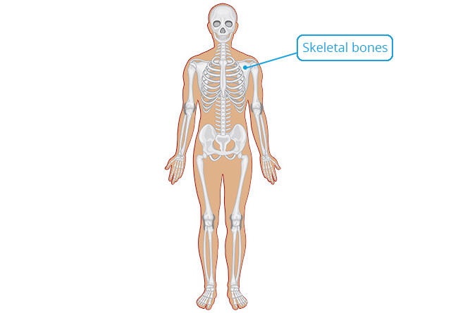
Bones.
Function
Provide great strength and support for the body; site of attachment of muscles; protect organs (e.g. skull protects brain); stores calcium.
Dense Irregular Tissue
Structure
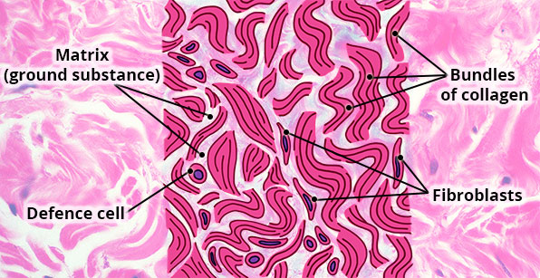
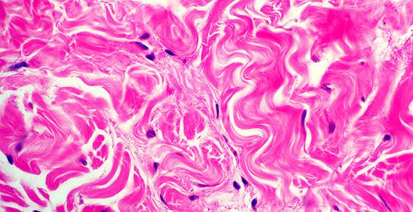
Bundles of densely packed collagen fibres running in many different directions. Fibroblast cells are located between the collagen bundles; very little open space (matrix) present.
Location
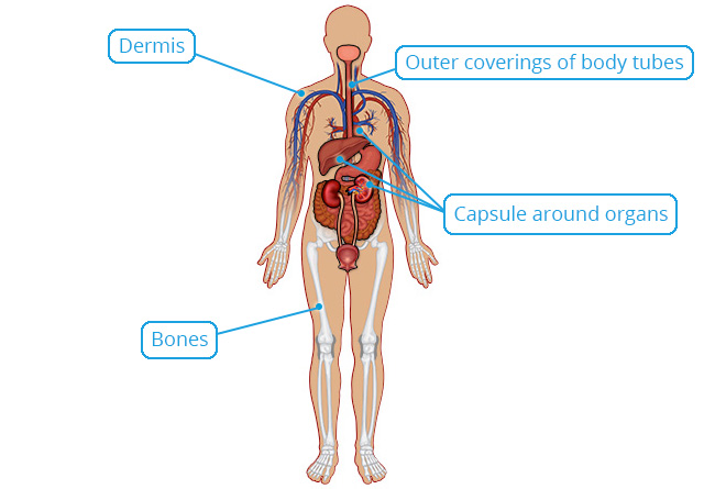
Beneath skin (dermis); capsules around organs (e.g. heart, kidneys, liver) and bones; outer coverings of body tubes e.g. oesophagus.
Function
Provides strength and resists stretching in all directions.
Dense Regular Tissue
Structure
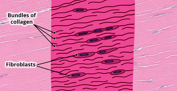
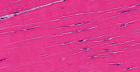
Bundles of densely packed collagen fibres running in the same direction; fibroblast cells located between the collagen bundles; very little open space (matrix) present.
Location
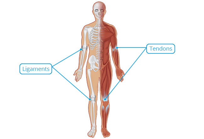
Forms tendons and ligaments.
Function
Can withstand strong pulling forces in the direction of the collagen bundles; tightly binds bone to bone (ligaments) and muscle to bone (tendons).
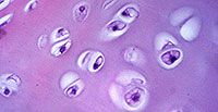
Hyaline Cartilage Tissue
Structure
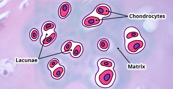
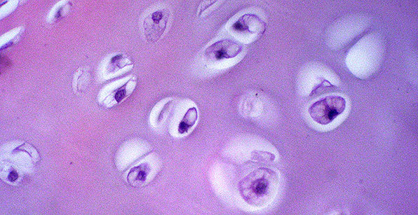
Clear matrix composed of very fine collagen fibres (not usually visible); many chondrocytes (cartilage cells) in clusters of 2-3 cells enclosed in a single space (lacunae).
Location
Cover ends of bones at moveable joints; in wall of trachea and bronchi; anterior ends of ribs; nose.
Function
Provides a smooth surface at joints that minimises friction and absorbs shock at joints; keep airways open; provides rigidity and flexibility in nose and ribs.
Loose (Areolar) Tissue

Structure
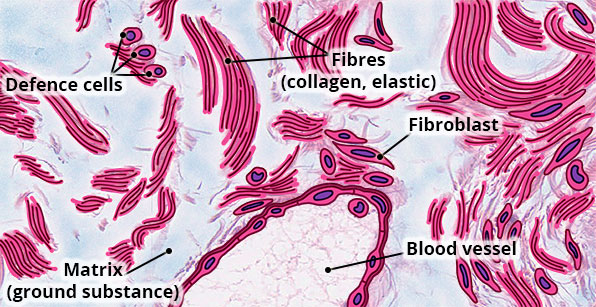
Loose arrangement of fibres (collagen, elastin, reticular), cells (fibroblasts, macrophages lymphocytes etc.) embedded in abundant ground substance.
Location
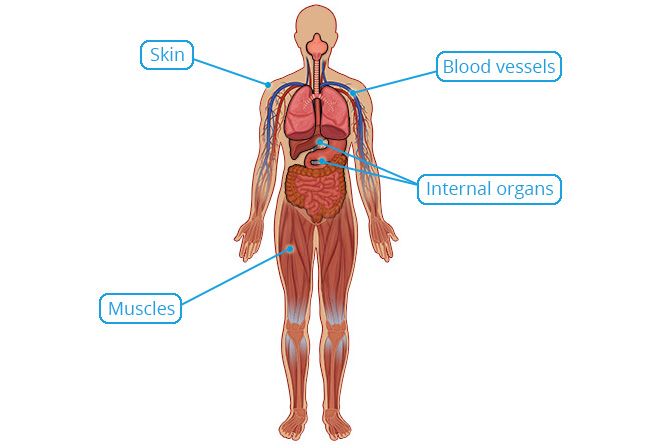
Widely dispersed throughout body; immediately beneath most membranous epithelial tissue, e.g. skin; around blood vessels, muscles and body organs.
Function
Loosely binds membranous epithelial tissue to deeper tissues; contains blood vessels to provide nutrients and oxygen to avascular epithelial tissue; support.
Reticular Tissue
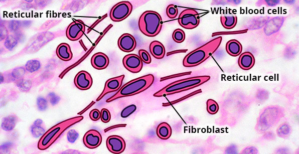
Structure
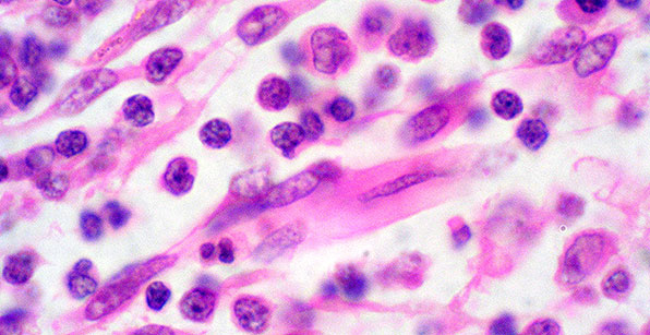
Loose network of reticular fibres and fibroblasts; the space between the fibres is filled with lymphocytes and other blood cells.
Location
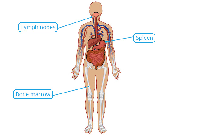
Within lymph nodes, the spleen and bone marrow.
Function
Forms a supporting structural framework; white blood cells remove old red blood cells in spleen and microbes in lymph nodes.
