The Cell : Structure
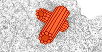

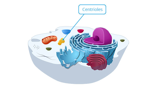
Centrioles
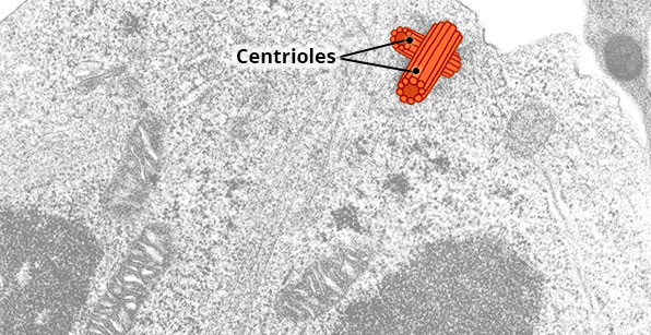

Location
Near the nucleus.
Appearance
A short, cylindrical structure composed of nine clusters of three rings of microtubules arranged in a circular pattern. A cell normally contains two centrioles which lie perpendicular to each other in a small area of the cytoplasm called the centrosome.
Function
The centrosome synthesises microtubules and plays an important role in cell division.
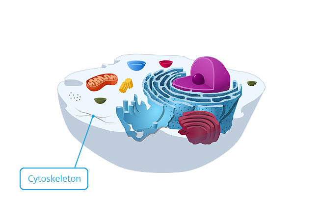
Cytoskeleton
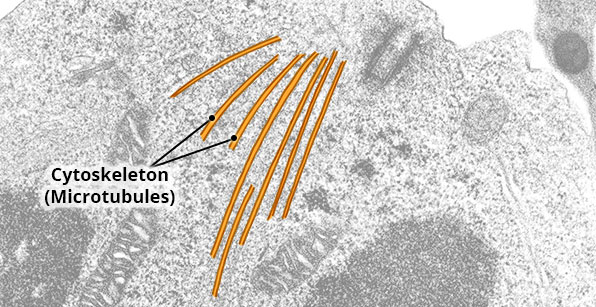
Location
Scattered throughout the cytoplasm.
Appearance
Network of three basic protein filaments: microfilaments, intermediate filaments and microtubules.
Function
Form the internal framework of a cell. Give a cell its strength, shape and support also aids in cell division.
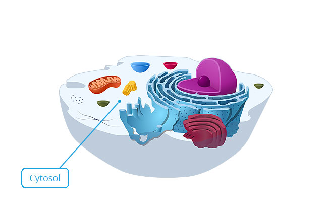
Cytosol
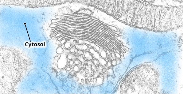
Location
Cytoplasm.
Appearance
Fluid that occupies at least 50% of a cell’s volume. Most of the cytosol is water, but proteins, sugars, amino acids and ions are also present.
Function
Where most of the chemical reactions occur in a cell.
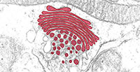
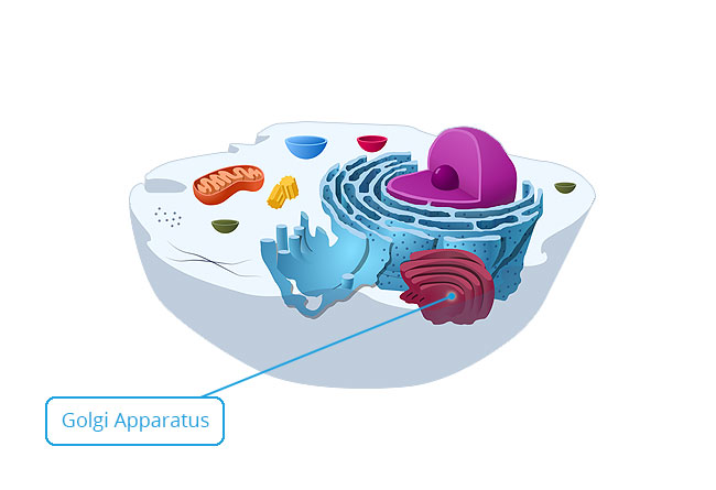
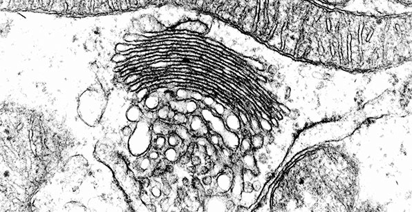
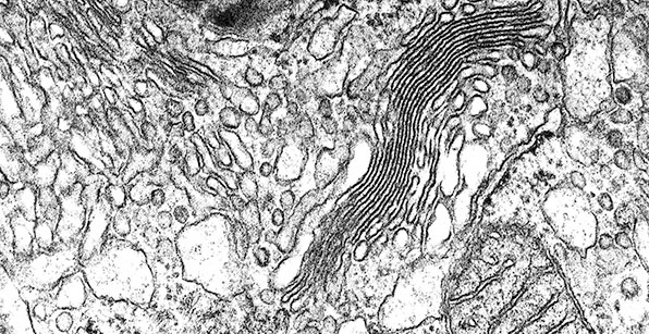
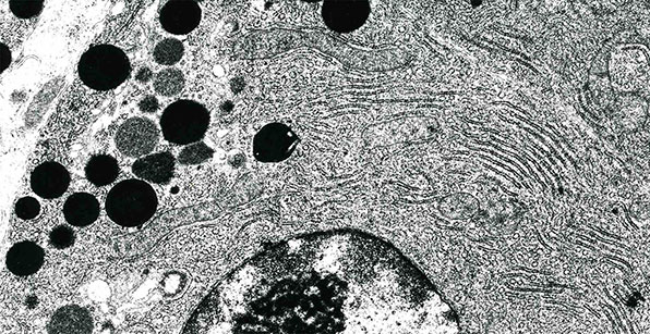
Golgi Apparatus
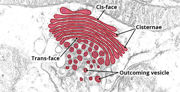

Location
Usually adjacent the nucleus and rough endoplasmic reticulum.
Appearance
A series of curved, flattened sacs called cisternae. The edges of each cisternae bulge out.
Function
The convex side of the Golgi apparatus (known as the entry or ‘cis’ face) receives newly synthesised proteins from the rough endoplasmic reticulum. The concave side (exit or ‘trans’ face) is where proteins leave the Golgi apparatus.
As proteins pass through the Golgi apparatus, they are concentrated and chemically modified by the addition of lipids and/or carbohydrate molecules. Modified proteins leave the Golgi apparatus via secretory vesicles.
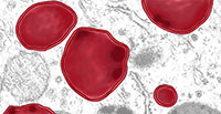
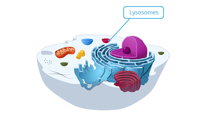
Lysosomes
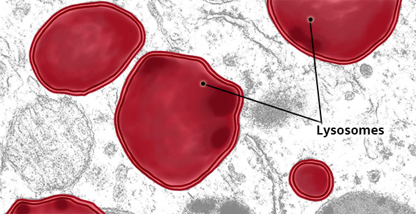
Location
Scattered throughout the cytoplasm.
Appearance
Vary in size and shape, but are membrane-bound organelles containing granular material. New lysosomes are round and pale, but older lysosomes tend to be irregularly shaped and electron-dense (i.e. dark in colour).
Function
Contain a variety of digestive enzymes that breakdown old and damaged cellular components, as well as ingested foreign material such as bacteria.
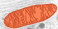
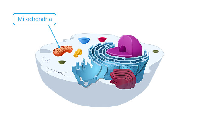
Mitochondria
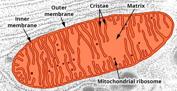

Location
Scattered throughout the cytoplasm, but localise where maximum energy is required.
Appearance
Vary in size and shape, but most often appear elongated. Consist of an outer permeable membrane, and an inner membrane that has folds called cristae. The inner membrane encloses a central, fluid-filled space called the matrix.
Function
The inner membrane is the site of production of most of a cell’s ATP, which is the energy source for the cell.
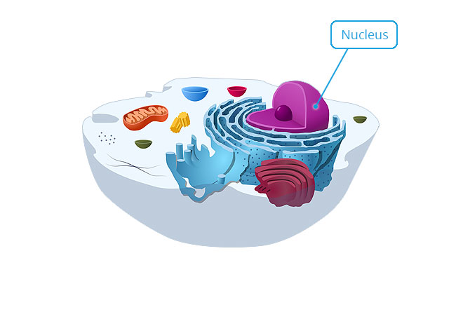
Nucleus
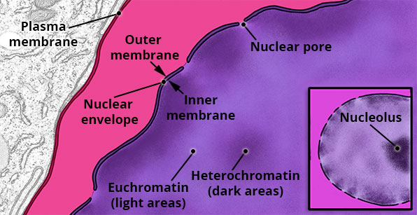
Location
Usually the centre of the cell.
Appearance
Spherical or oval-shaped. Red blood cells lack a nucleus, but osteoclasts (bone-dissolving cells) and skeletal muscle cells have many nuclei. The nucleus is surrounded by a double membrane called the nuclear envelope. The outer membrane of the nuclear envelope is continuous with the rough endoplasmic reticulum. The inner and outer nuclear envelope membranes regularly join to form nuclear pores.
The nucleoplasm contains fine threadlike matter called chromatin. Dense areas of chromatin are called heterochromatin and represent the portions of DNA that are not active in RNA (and thus protein) synthesis. Pale-staining euchromatin represents the part of DNA which is active in RNA/protein synthesis.
The nucleolus is a cluster of protein, DNA and RNA, and is the site of synthesis of ribosomal RNA (rRNA) and assembly of ribosomal subunits.
Function
Contains DNA that forms genes which direct all of the activities of the cell.
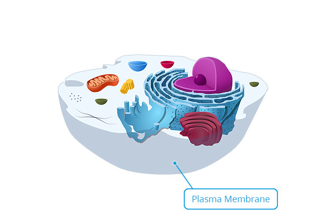
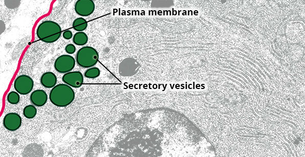
Plasma Membrane
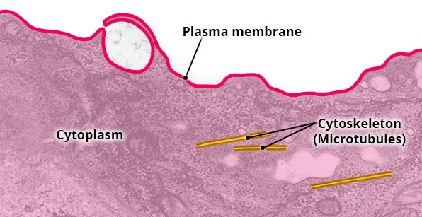
Location
External boundary of cell.
Appearance
Consists of phospholipid bilayer with embedded proteins.
Function
Regulates the movement of material into and out of the cell. Encloses cytoplasm.
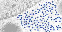
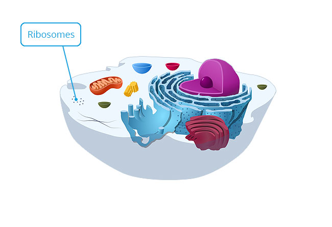
Ribosomes

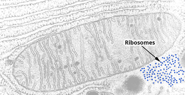
Location
Scattered throughout the cytoplasm.
Appearance
Small, round organelles. They can float around the cytoplasm as free ribosomes, or be attached to endoplasmic reticulum to form rough endoplasmic reticulum (RER).
Function
Site of synthesis of proteins for the cell’s own use.
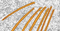
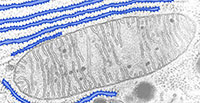
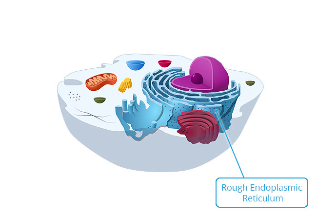
Rough Endoplasmic Reticulum
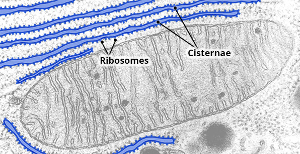
Location
Continuous with the outer membrane of the nuclear envelope.
Appearance
A series of parallel, flattened sacs (cisternae) studded with ribosomes.
Function
Site of synthesis of proteins for export, lysosomes and insertion into the plasma membrane.
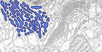
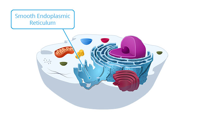
Smooth Endoplasmic Reticulum
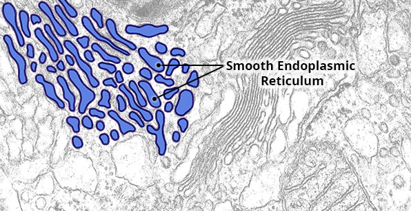
Location
Extend from the rough endoplasmic reticulum.
Appearance
An irregular network of flattened sacs. Unlike rough endoplasmic reticulum, no ribosomes are present on the outer surface of its membrane.
Function
Synthesises fatty acids, steroids like oestrogen and testosterone, and enzymes that detoxify alcohol and other drugs.
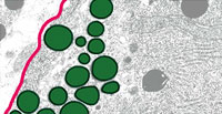
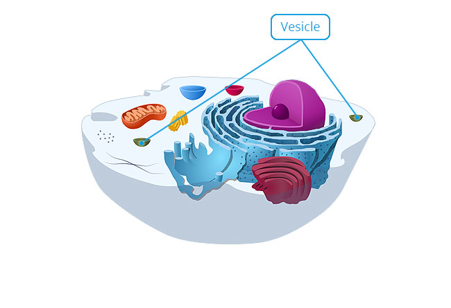
Secretory Vesicles
 <
<
Location
Accumulate between Golgi apparatus and plasma membrane where material is released from the cell.
Appearance
Membrane-bound. Pinch-off from the trans-face of the Golgi apparatus.
Function
Usually transport proteins from the Golgi apparatus to the exterior of the cell.